Advanced Imaging Techniques

Modern imaging technologies and advanced post-processing methods now allow epilepsy specialists to more precisely define the epileptogenic zone.

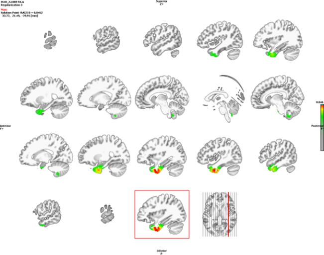
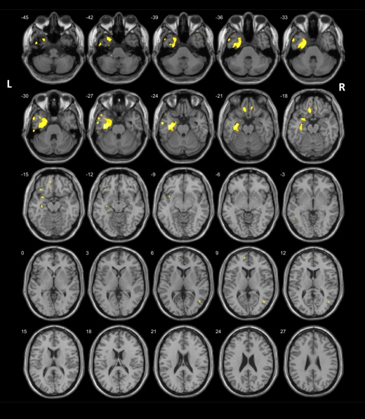
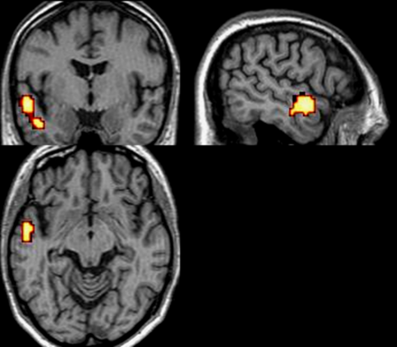
To confirm the hypothesis about the location of the seizure focus in the brain prior to surgery, a number of advanced diagnostic techniques can be employed. These methods are based on post-processing of data already acquired during previous imaging and diagnostic procedures.

They do not require the patient’s physical presence for new testing, but instead rely on the expertise of biomedical engineers, who use specialized computational tools to create enhanced visualizations. This allows for a more precise definition of the epileptogenic zone, supporting more accurate and tailored surgical planning.

ESI

Electric Source Imaging (ESI) combines high-density EEG (HD-EEG) recordings with the patient’s structural brain MRI to compute and visualize the three-dimensional distribution of electrical activity in the brain. The method is based on modeling the intracerebral electrical sources responsible for the EEG signals recorded on the scalp. Using complex mathematical algorithms and models, ESI estimates the location and orientation of these sources in relation to the individual’s brain anatomy.

ESI is particularly useful in pre-surgical assessment of patients with pharmacoresistant epilepsy, where accurate localization of the seizure onset zone is critical to a successful surgical outcome. Among all imaging modalities, ESI has shown the highest sensitivity and specificity in identifying the epileptogenic zone, outperforming conventional EEG, MRI, PET, and SPECT.

SPM PET

Statistical Parametric Mapping (SPM) is a method that compares a patient’s PET brain scan to a standardized database of scans from healthy controls, matched by age and sex.

By aligning the images to a common spatial template, SPM can statistically identify regions in the brain that show significant deviations from normal metabolic activity. This helps pinpoint areas potentially responsible for seizure generation and guides further treatment planning.

SISCOM

SISCOM (Subtraction Ictal SPECT Co-registered to MRI) is an advanced technique based on SPECT imaging that improves the precision of seizure focus localization. The process involves obtaining two SPECT scans: one during a seizure (ictal phase), one between seizures (interictal phase). 

These two scans are then subtracted from each other, and the resulting difference image is superimposed onto the patient’s brain MRI.

This allows clinicians to clearly identify areas of increased brain activity during the seizure, compared to the baseline interictal state. SISCOM provides critical functional information and helps localize the epileptogenic zone with high spatial accuracy.